Fig. 3.
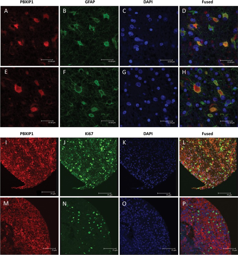
Immunofluorescence microscopy on paraffin-embedded sections of WHO grade IV glioblastoma multiforme. Two representative images of costaining with PBXIP1 (A and E; red), GFAP (B and F; green), and DAPI (C and G; blue) and the merged images (D and H) showing partial colocalization of PBXIP1 and GFAP expression. Two representative images of costaining with PBXIP1 (I and M; red), Ki67 (J and N; green), and DAPI (K and O; blue) and the merged images (L and P) showing PBXIP1 expression in Ki67-positive tumor area.

