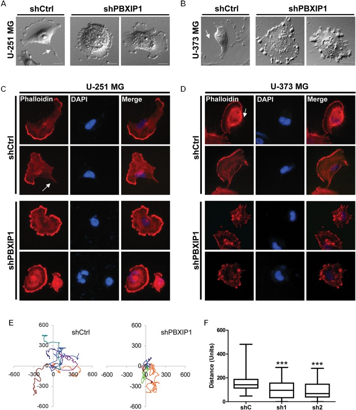
Fig. 6.
PBXIP1 depletion results in changes in cellular morphology and actin stress fiber formation and reduces motility of glioma cells. Representative bright-field images show the effect of loss of PBXIP1 expression in U-251 MG (A, right 2 panels) and U-373 MG HGG cells (B, right 2 panels) compared with the nontargeting shRNA control cells (A and B, left panels). Representative images of rhodamine-phalloidin staining in U-251 MG (C) and U-373 MG cells (D) with PBXIP1-knockdown (lower panels) or the nontargeting shRNA control cells (upper panels). Arrows indicate ventral actin stress fibers. PBXIP1-knockdown (E, right panel) impaired motility compared with the nontargeting shRNA control U-251 MG cells (E, left panel). Panel F shows the quantification of the distance over 24 h of the nontargeting shRNA control and the 2 independent shRNAs targeting PBXIP1 in U-251 MG cells. At least 50 cells were tracked. Distance is measured in arbitrary units. Plot F shows the mean, min, and max whiskers. The difference in motility with the control was significant for both shRNAs (P < .001).

