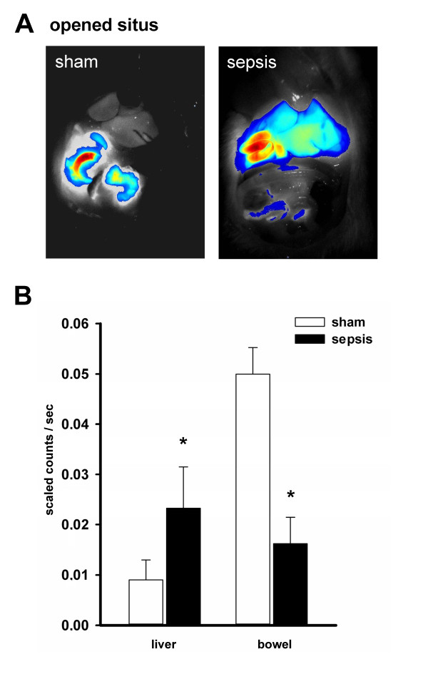
Figure 6.

Whole-body NIRF imaging at the opened situs clearly demonstrates an impairment specifically at the canalicular pole under conditions of sepsis. (A) Depicted are representative images taken 300 minutes after the experiment at the opened situs confirming an accumulation of ICG in the liver of septic animals, with an almost absent fluorescence signal in the bowel. The overlay is of fluorescence (false colors: blue (low fluorescence intensity) and red (high fluorescence intensity)) and white-light images. (B) Depicts the detected fluorescence intensities in liver and bowel of all animals at the open situs 300 minutes after administration of ICG. *P < 0.05 for sham versus sepsis ('liver': P = 0.008; 'bowel': P = 0.002).
