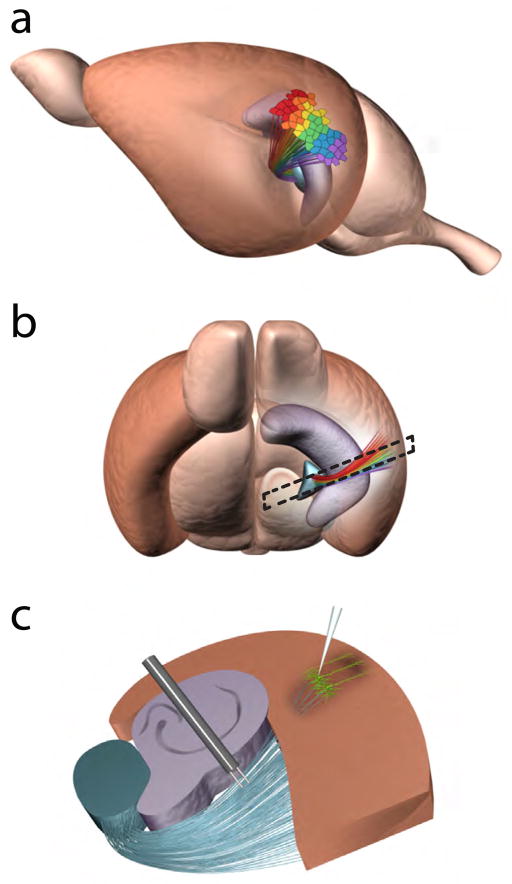
Figure 1. Thalamocortical projections form cortical maps in the primary ACx in mice.
(a, b) Cortical maps of sound frequencies in the primary ACx (brown region). The teal structure is the auditory thalamus (the ventral part of the medial geniculate, MGv) sending thalamocortical projections to the ACx, and the purple structure is the hippocampus. Different colors represent the tonotopy of thalamic projections and the primary ACx. Dashed lines in (b) represent the orientation and shape of the TC slice. (c) TC slice containing portions of the auditory thalamus, hippocampus, and ACx. LIII/IV pyramidal neurons (green) are the main thalamorecipient neurons in the primary ACx. Stimulating and recording electrodes are placed at the thalamic radiation and ACx, respectively. Note, the thalamic radiation contains multiple ascending and descending projections but only ascending thalamocortical projections are shown.

