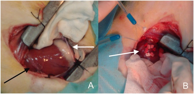
Figure 2. Views of the surgical approach.
A The thoracic cavity was opened via the right anterolateral thoracotomy at the fourth intercostal space, and the right ventricular anterior wall and apex were exposed. Black arrow showing the left anterior descending coronary artery (LAD). White arrow showing the right atrioventricular groove. B Two felt strip-buttressed purse-string sutures were placed at the right ventricular apex almost 10 mm far away from LAD (white arrow).

