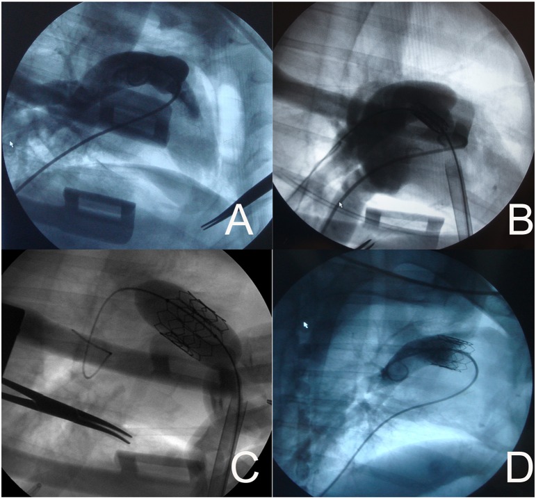
Figure 3. Valved stent implantation process (representative images).
A Imaging of the pulmonary valve was performed to measure the pulmonary valve radius and location. B After the valved stent was advanced to the pulmonary valve via the 22F sheath, a right ventricular angiography was performed to confirm that the valved stent was at the optimal position. C The valved stent was fully balloon-expanded. D A pulmonary angiography showing correct position of the valved stent in the pulmonary position in an sheep model. No regurgitation was assessed.

