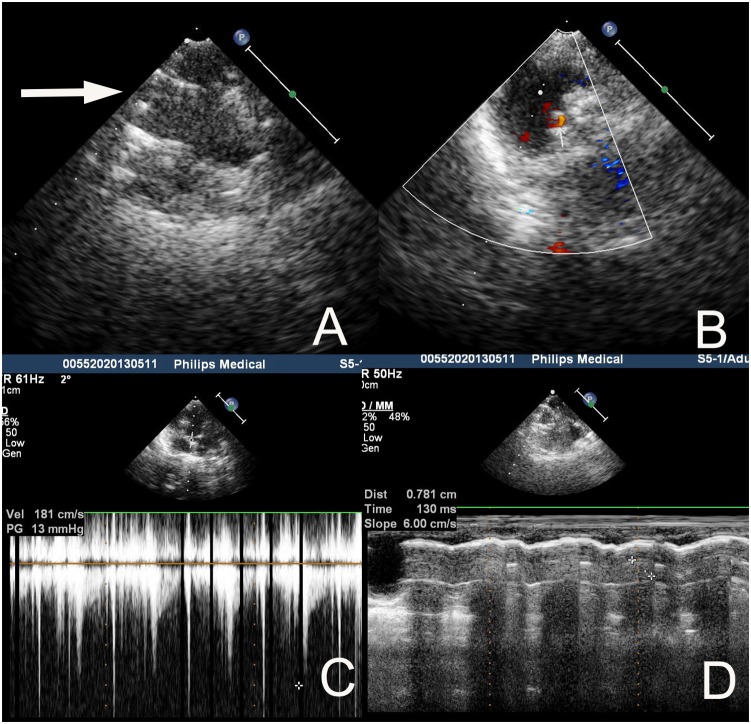
Figure 5. Echocardiography 4 weeks after implantation from a representative. sheep with a 20 mm PTPV.
A The two parallel lines of echo enhancement showed appropriate position and open shape of the stent (white arrow). B Color Doppler ultrasonography revealed no regurgitation or paravalvular leakage. C Doppler ultrasonography revealed the peak-peak transvalvular pressure gradient of the stented valve was 13 mmHg. D The motion distance of the valve cusp was 0.781 cm measured by Doppler ultrasonography, meaning normal function of the valve leaflet.

