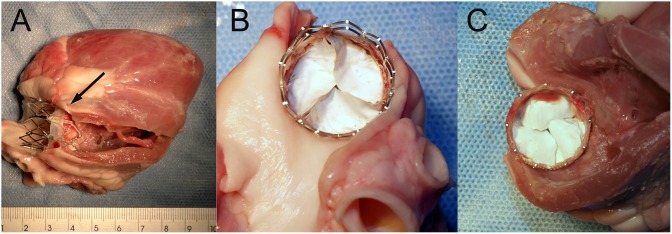
Figure 8. Gross morphology of the ePTFE valved stent explanted from a sheep 4 weeks after surgery.

A The native pulmonary valve can be seen (arrow), confirming the correct position of the valved stent. B The outflow side of ePTFE valved stent showed the leaflets were thin without significant tissue deposits. C The inflow side of ePTFE valved stent showed slight fibrous overgrowth at the bottom of the leaflets, in the commissural areas and on the sealing cuff.
