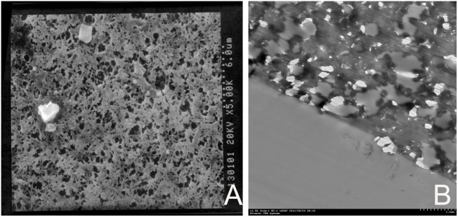
Figure 10. Electron microscopic images of the ePTFE leaflets explanted from a sheep 4 weeks after surgery.

A Scanning electron microscopic appearance of the outflow side of a ePTFE valve cusp showing polyporous structure of ePTFE and no obvious cell or tissue attachment (×5000). B Transmission electron microscopic appearance of the outflow side of a ePTFE valve cusp showing polyporous structure of ePTFE and no obvious cell or tissue infiltration. (×1500).
