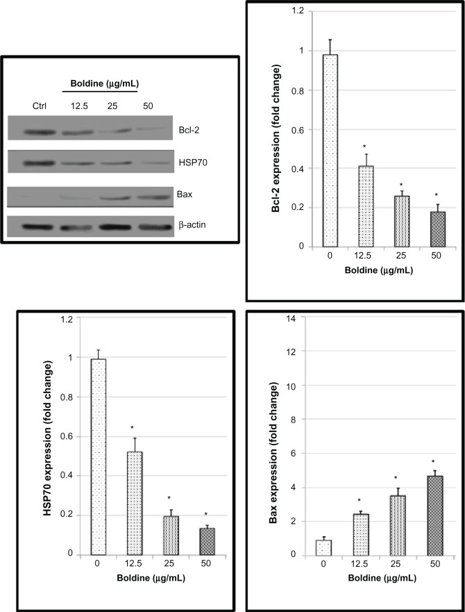
Figure 9.
Western blot analysis of boldine-treated MDA-MB-231 cells. Cells were treated with boldine for 24 hours before being lysed and subjected to separation by sodium dodecyl sulfate polyacrylamide gel electrophoresis. Proteins were then transferred to membrane and probed with antibodies against Bax, Bcl-2, and heat shock protein 70. The membrane was reprobed with anti-β-actin antibody as the loading control. The band densities of the boldine-treated samples were normalized to the control. Data are shown as the mean ± standard deviation (n=3). Data were analyzed by the Student’s t-test (*P<0.05).

