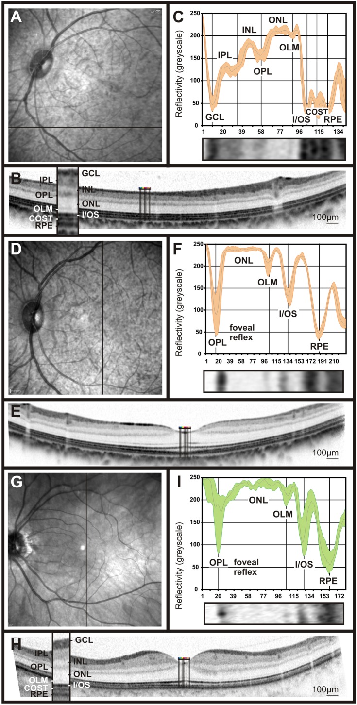
Figure 4. Retinal imaging and OCT reflectivity profile in cynomolgus monkeys and comparison to humans.
SLO native fundus imaging in non-human primates (A, D) and one of the researcher’s eye (G). The solid bar indicates the origin of the OCT scan. A representative OCT scan taken from the ventral retina (B), the fovea of the non-human primate (F) and the researcher’s eye, (H). OCT reflectivity profiles from B, F and H were extracted and assigned to the retinal layers of cynomolgus monkeys fovea and extrafoveal regions (F and C, respectively) and the human fovea (I).

