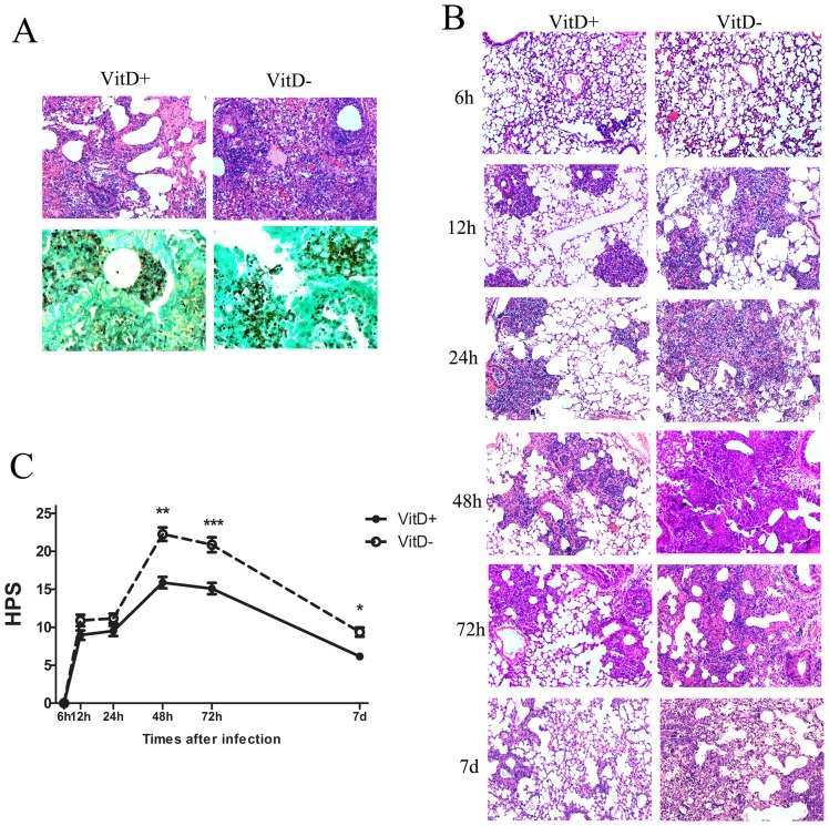
Figure 2. Histological evidence of aggravated and sustained inflammation in VitD- mice challenged with A. fumigatus conidia.
A. Representative lung sections from VitD+ and VitD- mice three days post intratracheal challenge with 5×10⁷ A. fumigatus conidia. Sections were stained with HE (upper, magnification 100x) for analysis of inflammation, and with GMS (lower, magnification 400x) for the detection of conidia and hyphae. B. Representative HE-stained lung sections (magnification 100x) from VitD- and VitD+ mice are shown at the indicated times after intratracheal challenge with 2×10⁷ A. fumigatus conidia. C. HPS of serial HE-stained lung sections between 6 h and 7 d after intratracheal challenge with 2×10⁷ A. fumigatus conidia. HPS is presented as mean±SE. Data are representative of three independent experiments (n = 3/group). Statistical analysis was performed by a Mann-Whitney rank sum test. p<0.001(***), p<0.01(**), p<0.05(*).

