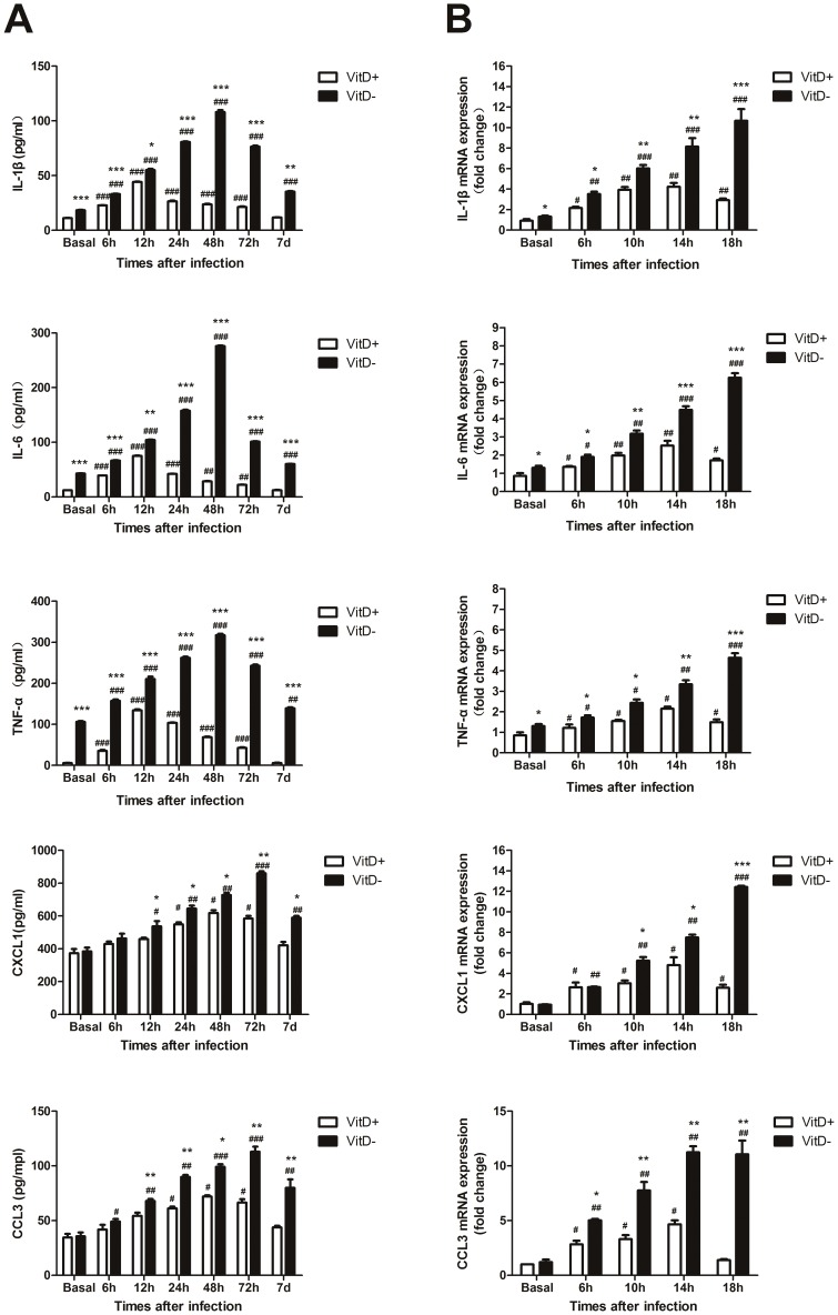
Figure 4. Enhanced pro-inflammatory cytokine and chemokine production in the lungs of A. fumigatus-challenged VitD- mice.
A. Mice were infected intratracheally with 2×10⁷ A. fumigatus conidia. At specific times p.i. BALF collected from mice was analyzed for cytokine and chemokine content by ELISA. Data are representative of three independent experiments with n = 5 per group (mean±SE). p<0.001(###), p<0.01(##), p<0.05(#), for comparison of A. fumigatus-challenged VitD+/VitD- mice versus baseline levels; p<0.001(***), p<0.01(**), p<0.05(*), for comparison of VitD+ with VitD- mice using paired Student's T-test. B. AMs isolated from naïve mouse lungs by BAL, were incubated in medium alone or in the presence of viable A. fumigatus conidia (MOI = 0.1) in vitro. Total RNA from AMs was extracted at indicated times. Transcript abundance was measured by real-time RT-PCR. Representative data from three independent experiments with n = 3 per group are shown. P<0.001(###), P<0.01(##), P<0.05(#), for comparison of A. fumigatus-challenged VitD+/VitD- AMs versus basal levels; P<0.001(***), P<0.01(**), P<0.05(*), for comparison of VitD+ and VitD- AMs using paired Student's T-test.

