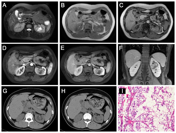
Figure 4. Translocation RCC in a 36-year-old woman (patient 9 in Table 1, 2, 3) with gross hematuria.
A–C, Axial T2WI and plain (non-contrast) T1WI (B, IP in phase; C, OP) showing an irregular well-defined mass (T2, iso-high mixed signal intensity; T1, slightly high signal intensity) with a patchy area of cystic necrosis in the left renal pelvis. Axial CMP (D) and NP (E) gradient-echo MR images showing moderate heterogeneous enhancement in the left renal mass. F, Coronal delay phase image showing a delayed-enhancement mass within the contour of the kidney. G–H, Unenhanced CT images showing a well-defined, slightly high-density renal mass with mottling calcification. I, The tumor cells contain hyaline cytoplasm with a papillary structure (HE 10 & 40).

