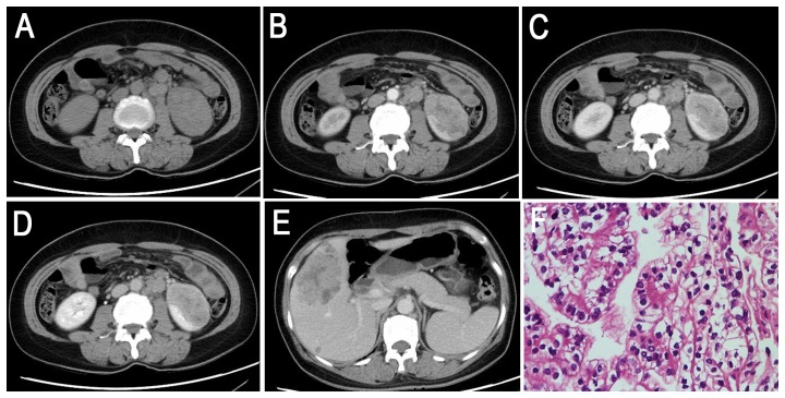
Figure 5. Translocation RCC in a 40-year-old woman (patient 10 in Tables 1, 2, 3) complaining of left flank pain for approximately half a year that became more serious 1 month prior to imaging.
A, Axial unenhanced CT image showing an ill-defined, slightly higher attenuation soft tissue mass in the left kidney. Retroperitoneal adenopathy is present. B–D, Axial CMP, EP and NP contrast-enhanced CT images demonstrating moderate and prolonged enhancement of the mass without an integrated capsule. E, Liver metastasis with hemorrhage on EP is observed. F, The tumor cells consist of poorly differentiated clear cells (HE 10 & 40).

