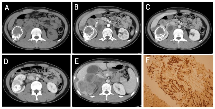
Figure 6. A 16-year-old boy (patient 12 in Tables 1, 2, 3) with XP11.2/TFE 3 confirmed by biopsy.
A, Axial unenhanced CT image showing an ill-defined, irregular, slightly higher attenuation mass with a bulk of plaque-like calcifications in the right kidney. Retroperitoneal adenopathy is observed. B–D, Axial CMP and NP contrast-enhanced CT showing a heterogeneously enhanced mass. E, The liver and porta hepatis areas and right retroperitoneal multiple lymphoma metastases are indicated. F, Immunohistochemical analysis demonstrated TFE3 nuclear staining (HE 20 & 10).

