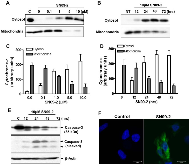
Figure 6. SN09-2 stimulates cytochrome c release from mitochondria and caspase-3 activation.
(A–D) Western blotting of cytochrome c in the cytosolic and mitochondrial fractions (A, C) PC3 cells were treated with different concentrations of SN09-2 for 24 h and fractionated into cytosol and mitochondria. 20 µg lysates from each fraction were used for SDS-PAGE and western blotting with anti-cytochrome c antibodies. (B, D) Cells treated with 10 µM SN09-2 were harvested at different time points, and then fractionated. (C, D) The signal intensity of each blot was measured and shown as graphs (E) PC3 cells treated with 10 µM SN09-2 were harvested at different time points, and used for western blotting with anti-caspase-3 or a cleaved form of caspase-3 antibodies. The amount of loaded proteins was normalized with anti-β-actin antibodies. (F) SN09-2-treated PC3 cells on cover slips were labeled with antibodies for active cleaved caspase-3 and anti-β-actin, and then briefly stained with Hoechst33342. Fluorescence signals were observed under a confocal microscope.

