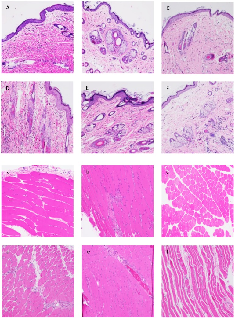
Figure 1. Histological evaluation of skin and muscle rejection in the early postoperative phase (POD 3 and 5).
(A–C) Skin sample taken on POD 3 from an allograft without immunosuppression (A), from an isograft (B) and an allograft under TAC (C) showing no/rare inflammatory response (Grade 0 rejection). (D–F) Skin biopsies taken on POD 5 from a rejecting animal (D) displaying Grade 1 rejection, from an isograft (E) showing no/rare inflammatory response and a TAC treated allograft (F) characterized by a mild inflammatory response (Grade 0-I) in the deep dermis. (a–c) Muscle sample taken on POD 3 from an allograft without immunosuppression (a), from an isograft (b) and an allograft under TAC (c) showing no/rare inflammatory response (Grade 0 rejection). (d–f) Skin biopsies taken on POD 5 from a rejecting animal (d) displaying a mild inflammatory response (Grad 0-I rejection), from an isograft (E) and a TAC treated allograft (f) showing no/rare inflammatory response.

