
Figure 8.
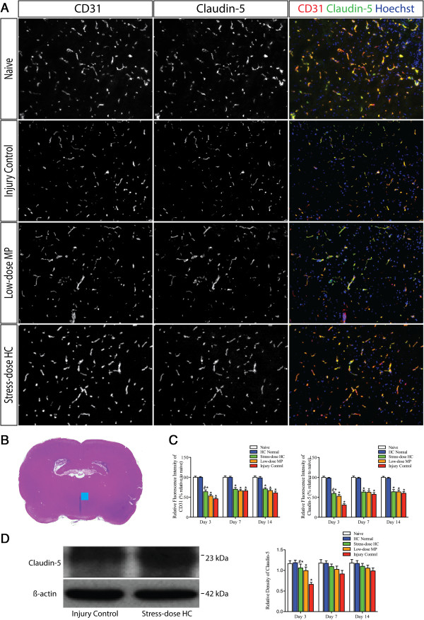
Stress-dose hydrocortisone reduces the loss of CD31 and claudin-5 in the paraventricular nuclei (PVN) of the hypothalamus following trauamatic brain injury (TBI). (A) Representative images of blood vessels in ipsilateral hypothalamus, immunostained for endothelial cell marker CD31, and tight junction protein Claudin-5. Stress-dose hydrocortisone (HC) appears to partially preserve loss of CD31 and claudin-5 caused by TBI, more pronounced with claudin-5. (B) Region of interest for immunoreactivity analysis was the PVN of the bilateral hypothalamus. Blue square indicates where the immunofluorescence pictures (A) were taken. (C) Summary data shows immunoreactivity of CD31 and claudin-5 were lowest on post-injury day 3, when significant BBB leakage was detected by EB extravasation measurement. Importantly, they were significantly increased by the administration of stress-dose HC, when overt improvement of BBB permeability was also observed. Analysis of variance (ANOVA), F(4,45) = 54.625, P = 0.000 for CD31 comparison on day 3; post hoc least significant difference (LSD), P = 0.001 for stress-dose HC versus injury control, P = 0.000 for injury control versus naïve. F(4,45) = 82.263, P = 0.000 for claudin-5 comparison; post hoc Dunnett T3, P = 0.000 for stress-dose HC versus injury control, P = 0.000 for injury control versus naive. (D) Representative western blot and summary data shows increased claudin-5 expression in the hypothalamus following administration of stress-dose HC on post-injury day 3. ANOVA, F(4,31) = 5.716, P = 0.001; post hoc LSD, P = 0.001 for stress-dose HC versus injury control, P = 0.003 for injury control versus naive. At each time point: n = 3 from each of the two groups (naïve and HC normal), n = 10 from each of three groups (injury control, low-dose methylprednisolone (MP), and stress-dose HC). Data are mean ± standard error of the mean.