
Fig. 3. YAP1 deletions and expression in MM cell lines and samples from subjects with MM.
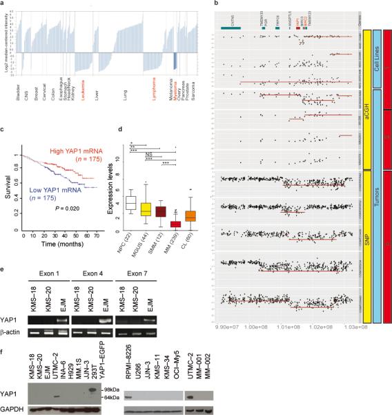
(a) YAP1 mRNA expression in solid tumors and hematological cancers (Oncomine at www.oncomine.org; Wooster Cell Line dataset).
(b) Gene dosage comparison across cell lines and representative tumors demonstrating homozygous deletions at the YAP1 locus. Number 1 corresponds to data derived from 21, 2 from 23, and 3 from 24.
(c) Survival curve relative to YAP1 expression in individuals affected by MM, obtained from www.canevolve.org, and based on GSE2658.
(d) Expression data comparing plasma cells from healthy subjects, MGUS, MM, and cell lines (CL), combining data from GSE5900, GSE2658, and from the MMRC collection (http://www.broadinstitute.org/mmgp), probe set 224895_at. *: P < 0.05; **: P < 0.01; ***: P < 0.001, one-way Anova, Dunn's Multiple Comparison Test.
(e) Non–quantitative PCR on genomic DNA from YAP1–deleted MM cell lines KMS–18 and KMS–20 and control cell line (EJM).
(f) Western blot analysis of YAP1 in MM cell lines and cells derived from subjects with MM. As positive control, lysates from 293T cells transfected with YAP1–EGFP vector were used (band at 98 kDa).