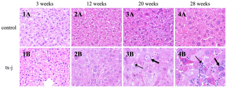
Figure 3.
Histology images of tx-j and control livers from three to 28 weeks of age. All hematoxylin and eosin stained, 436×. Whereas liver histology was normal in tx-j mice at both three and 12 weeks of age (1B and 2B), there was an increase in inflammatory infiltrates at 20 and 28 weeks (3B and 4B, thin arrows), in association with giant nuclei and markedly increased cell size of the hepatocytes (3B and 4B, thick arrows). Note that in tx-j mice hepatocyte cell diameters are 2.5 times and nuclear diameters are about two times larger than control mice [19]. Control mice had normal liver histology at all time points (1A, 2A, 3A and 4A).

