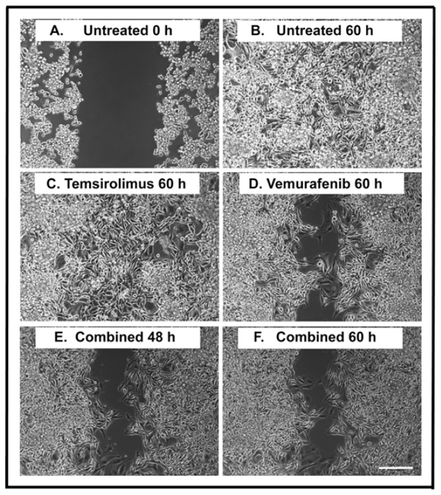
Figure 2.
Wound healing assay of H1_DL2 melanoma brain metastasis cells after treatment with vemurafenib and temsirolimus. (A) Untreated H1_DL2 cells at 0 h; (B) Untreated cells 60 h after initiating a scratch wound; (C) Representative picture 60 h after initiating a scratch wound and starting the treatment with 5 μM of temsirolimus; (D) Representative picture 60 h after initiating a scratch wound and starting the treatment with 5 μM of vemurafenib; (E) Representative picture 48 h after initiating a scratch wound and starting combined treatment (5 μM temsirolimus and 5 μM vemurafenib); and (F) Representative picture 60 h after initiating a scratch wound and starting combined treatment (5 μM temsirolimus and 5 μM vemurafenib). Scale bar = 200 μm.

