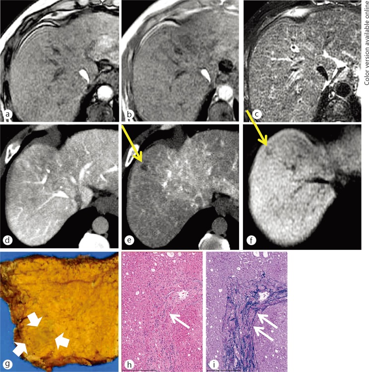
Fig. 2.
A 50-year-old man with an early HCC. There was no nodule on the opposed-phase (a), the inphase (b) T1-weighted MR images or the T2-weighted MR image (c). There was no nodule on CT image during arterioportography (d) because the nodule showed equivalent portal flow compared with the surrounding liver parenchyma. The nodule (yellow arrows) showed decreased arterial flow on CT imaging during hepatic arteriography (e). The nodule was detected as hypointense on hepatocyte imaging via EOB-MRI (f). On histological examination, this nodule was identified as a small nodule with an unclear margin (thick white arrows) macroscopically (g). Portal invasion (white short arrows), which is a unique finding to early HCC, was identified on hematoxylin and eosin staining (h) and Victoria-blue staining (i), respectively.

