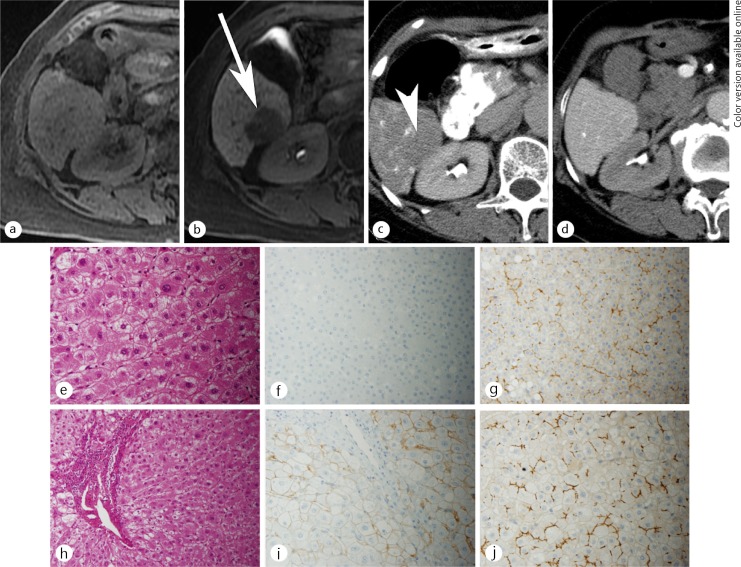
Fig. 6.
A 58-year-old man with an early HCC. No lesion was shown on the unenhanced T1WI (a). Hepatocyte phase imaging (b) showed a well-defined nodule at the posterior segment of the right lobe of the liver (arrow). The nodule showed decreased uptake of gadexetic acid (relative enhancement ratio of 0.52), resulting in hypointensity relative to the surrounding liver. This nodule showed a slight decrease of arterial flow on CT imaging during hepatic arteriography (c), and equivalent of portal venous flow during arterioportography (d) compared to the surrounding liver. The histological examination of the nodule (e-g) compared with the surrounding liver (h-j), showed slight cellular atypia and mild increased cell density with thick trabecular pattern on hematoxylin and eosin stain (e), no expression of OATP1B3 (f), and equivalent expression of MRP2 (g) compared with surrounding liver (h, i, j, respectively).

