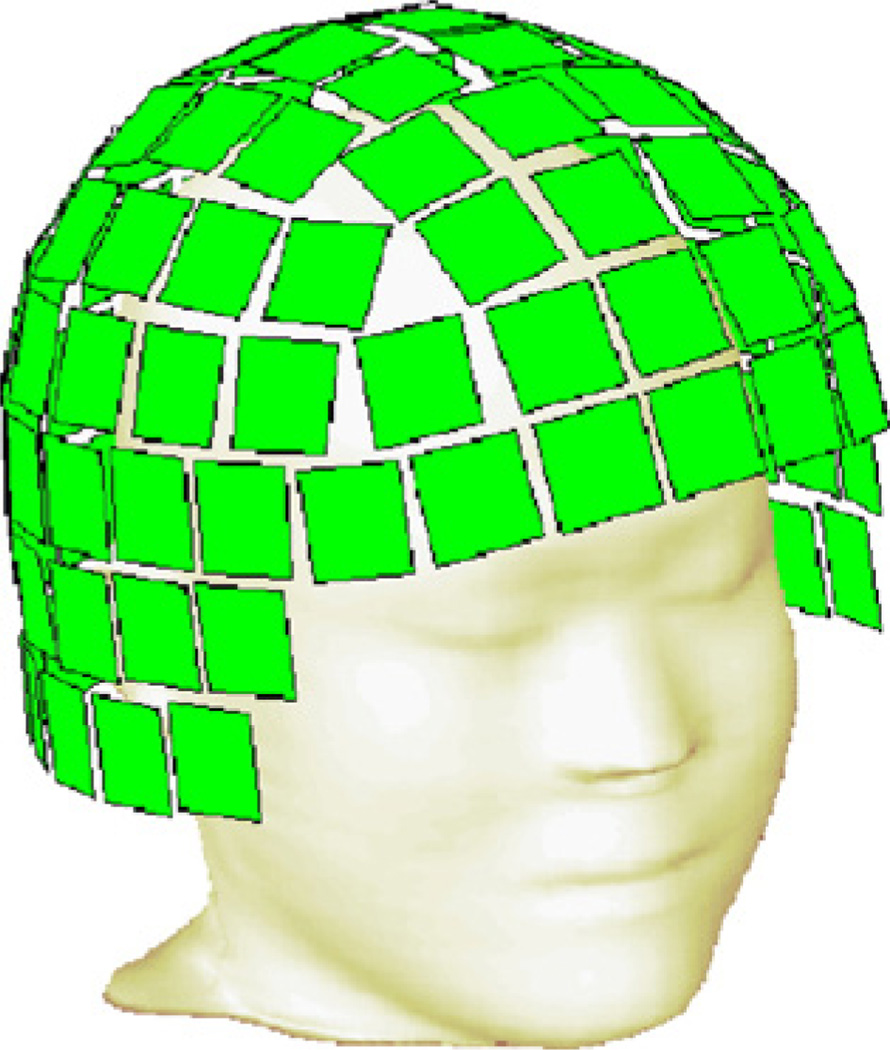
Fig. 3.
Geometry that was used in the model study. An outer head surface was extracted from the subject's MRI. MEG sensor array (a “hat” consisting of square receiver “plates”, as shown here) was positioned in MRI coordinates by matching the reference points to the head surface. Each “plate” measures normal component of a magnetic field.

