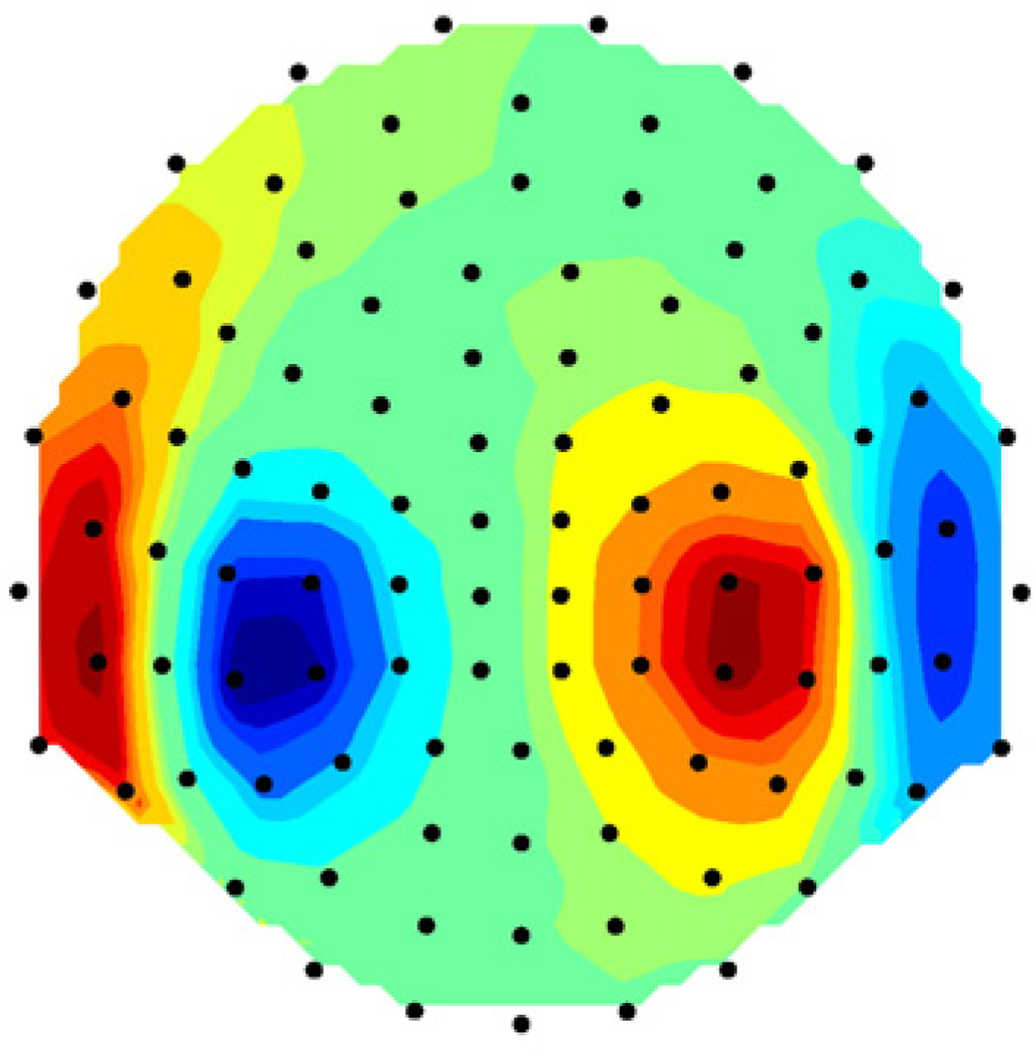
Fig. 5.
Magnetic field data for two-dipole model (the model from Fig. 4). Data are shown by color map superimposed on flat projection of measuring array (the helmet from Fig. 3). Dots show the locations of the sensors, each sensor corresponds to one plate in Fig. 3. Data contain Gaussian random noise such that the SNR=400.

