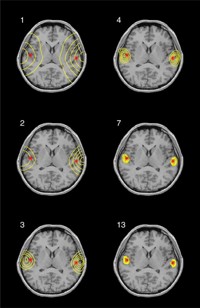
Fig. 7.
Evolution of solution during reweighted iterations. The case corresponds to example discussed in Fig. 6. Stars show “true” location of dipoles (location of dipoles within the head is shown in Fig. 4). Solution is superimposed on corresponding MRI slice as isolines. Panels numbered 1, 2, 3, 4, 7, 13 show the solutions at the corresponding iteration.

