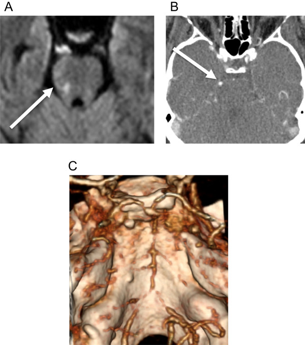
Figure 1.

Right pons ischemia. A. Diffusion weighted MRI showing restriction in right pons (arrow) consistent with ischemia on October 29, 2012. B. Contrast CT with right superior cerebellar artery (arrow) enhancement. C. Reformatted 3-D CT angiography on Oct 29, 2012 showing diffuse vasculopathy at the basilar arteries and bilateral vertebral arteries.
