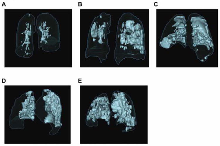
Figure 2. Extent of Radiographic Pulmonary Pathology at the Onset of Alveolar Hemorrhage.

(A-E) Representative surface renderings of pulmonary parenchymal pathology at the onset of alveolar hemorrhage are shown from five different patients treated with rFVIIa and conventional therapy. (A) Patient 8; 8%, (B) Patient 21; 20%, (C) Patient 5; 25%, (D) Patient 13; 37%, and (E) Patient 11; 47%. Total lung volume is outlined in blue and pathological areas are highlighted in light green.
