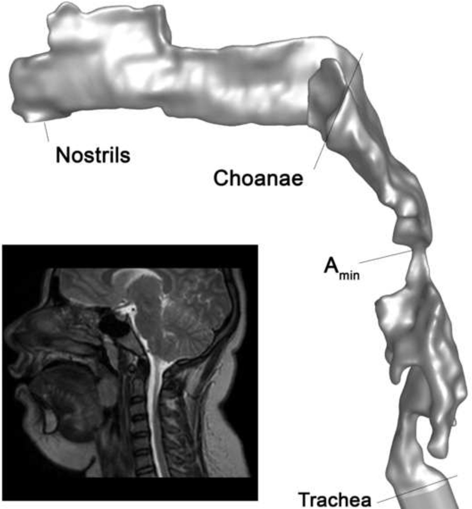
Figure 1.
3D upper airway model of subject 4 pre-surgery based on reconstructed segmented MR axial images. Anatomical locations along the airway model are shown for reference. Amin is the location of minimum cross-section where tonsils and adenoids constrict the pharynx; in this subject Amin is located between the tonsils in the retrolingual pharynx. Inset: midline sagittal MR image of subject 4.

