Figure 1.
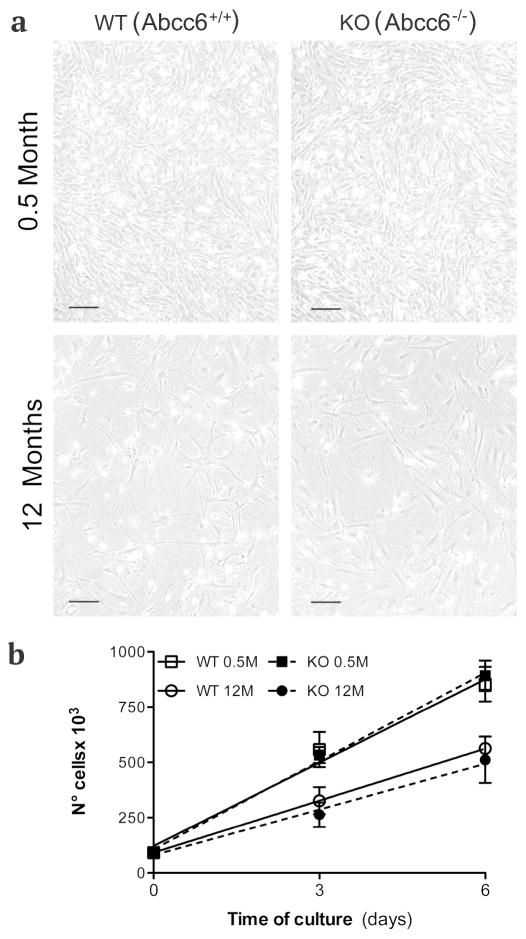
Dermal fibroblasts cultured from KO (Abcc6−/−) and WT (Abcc6+/+) mice of 0.5 and 12 months of age and observed by phase contrast microscopy (a). Differences in the proliferation capabilities are related to the age of the animals, but are independent of the genotype as clearly shown in graph (b). Bar = 300 μm.

