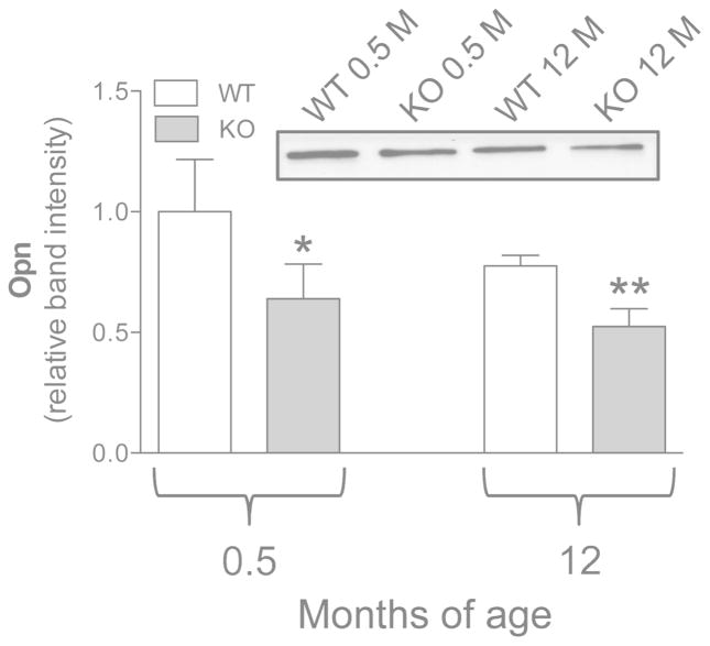
Figure 6.
Expression of Opn measured by Western blot in dermal fibroblasts cultured from WT (Abcc6+/+) and KO (Abcc6−/−) mice of 0.5 and 12 months of age. Data are expressed as mean values ± SD of densitometric analyses were values in cells from WT animals 0.5 month-old were set at 1. A representative Western blot is shown. *p<0.05 and **p<0.01 KO vs WT of the same age.

