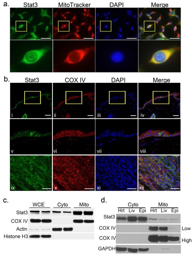
Figure 1. Stat3 in keratinocyte mitochondria.
(A) Mitochondrial localization of Stat3. Mouse 3PC keratinocyte cells were incubated with MitoTracker for 15 minutes, then permeabilized and stained for Stat3 (green). Scale bars = 50 μm
(B) Colocalization of Stat3 and mitochondrial marker COX IV in vivo. Mouse skin (i-viii) and SCC (ix-xii) sections were co-immunostained with mouse anti-Stat3 (green) and rabbit anti-COX IV (red). Scale bars = 50 μm.
(C) Detection of Stat3 in mitochondrial enriched fractions. Western blot analysis of mitochondrial fractions from primary culture adult mouse keratinocytes and (D) Analysis of mitoStat3 in vivo. Mitochondrial fractions from mouse heart (Hrt), liver (Liv) and skin epidermal cells (Epi). Actin and GAPDH were used as cytoplasmic markers, while COX IV was used as mitochondrial marker [lighter (low) and darker (high) exposures are shown]. WCE; whole cell extract, Cyto; cytoplasmic fraction, Mito; mitochondrial fraction.

