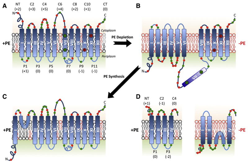
Fig. 3.
Topological organization of LacY, PheP, and GabP as a function of membrane lipid composition. TMDs (Roman numerals) and EMDs (Arabic numerals) are sequentially numbered from the N-terminus to C-terminus with EMDs exposed to the periplasm (P) or cytoplasm (C) as in wild type cells. Net charge of EMDs and distribution of positive (red) and negative (green) charges are shown. Topology of LacY is shown after initial assembly in PE-containing cells (A), after either initial assembly in PE-lacking cells or after dilution of PE following initial assembly in PE-containing cells (B), or after initiation of PE synthesis post-assembly of LacY in PE-lacking cells (C). EMD P7 is recognized by monoclonal antibody 4B1 in PE-containing (specific conformation) but not in PE-lacking (loss of native conformation) cells. Charges in TMD VII salt bridge with charges in TMDs X and XI in native LacY. The interconversion of topological conformers (A, B and C) is reversible in both directions. Topology and EMD charge distribution of the lipid sensitive domains of PheP and GabP are shown in PE-containing and PE-lacking cells (D).

