
Figure 2.
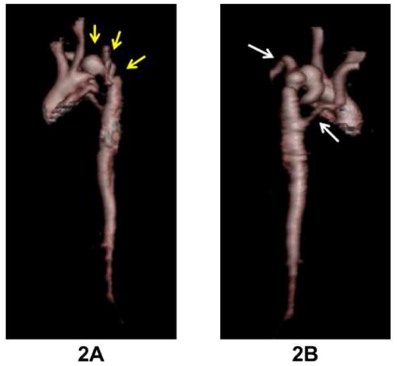
Three dimensional MRI aortic reconstruction in this PHACE patient with severe coarctation with multiple areas of narrowing and aneurysms in the transverse arch (as outlined by arrows in 2A). Both subclavian arteries arise distal to areas of obstruction (arrows in 2B), and the right subclavian artery has an aberrant origin.