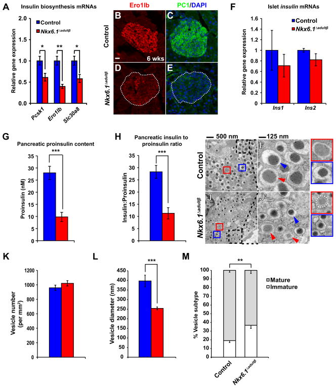
Figure 5. Nkx6.1 is necessary for insulin biosynthesis.
(A) qRT-PCR analysis of islets reveals reduced expression of genes involved in insulin biosynthesis in Nkx6.1 Δadultβ compared to control mice at 6 wks (n=3). (B–E) Immunofluorescence staining of pancreata from Nkx6.1 Δadultβ and control mice at 6 wks. Dashed lines represent islet area. (F) qRT-PCR analysis of Nkx6.1 Δadultβ and control islets from mice at 6 wks shows no significant difference in ins1 or ins2 expression (n=3). (G) Proinsulin content normalized to protein concentration of whole pancreatic lysates (n=6). (H) The pancreatic insulin to proinsulin ratio is reduced in Nkx6.1Δ adultβ mice (n=6). (I,J) Transmission electron microscopy of pancreatic sections reveals smaller vesicle size and an increase in immature vesicles (red arrowheads) in Nkx6.1 Δadultβ compared to control mice. Dashed boxes indicate area of magnification in I′ and J′. Blue arrowheads point to vesicles containing mature insulin dense core granules. Insets framed red show a representation of a typical immature vesicle and insets framed blue a typical mature vesicle. (K–M) Quantification of vesicle numbers (K), vesicle diameter (L), and the percentage of mature and immature vesicles (M) in Nkx6.1Δ adultβ and control mice (n=10). In G–M, mice were analyzed at 7 wks. PC1, prohormone convertase 1/3; Wk, week. Data are shown as mean ± SEM for A,F,G,H and ± SD for K–M. *p<0.05, **p<0.01, ***p<0.001.

