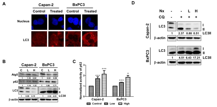
Figure 1. Nx treatment inhibits autophagy in human pancreatic cancer cell lines.
A. Logarithmically growing human pancreatic cancer cells Capan-2 and BxPC-3 treated with or without Nx (150 μg/ml for Capan-2 and 60 μg/ml for BxPC-3) for 24h were used to determine LC3 levels using immunofluorescence microscopy. Experiment was repeated three times and a representative picture is shown. B. Whole cell extracts prepared from logarithmically growing Capan-2 and BxPC-3 cells treated with low and high dose Nx (doses shown in table 1) for 24h were used in immunoblot analysis with Atg5, p62 and LC3. The membrane was probed with β-actin for loading control. Quantification data normalized to -actin were shown below the blot. Representative blot from multiple experiments is shown. C. Logarithmically growing Capan-2 and BxPC-3 cells treated with low and high Nx for 24h were used to determine the levels of p62 using ELISA (Enzo life sciences, NY). Statistical significance between groups was determined using students t-test and p values less than 0.05 was considered significant (* p<0.05, and *** p<0.005). D. Whole cell extracts prepared from logarithmically growing Capan-2 and BxPC-3 cells treated with different doses of Nx (low and high) in the presence and absence of autophagy inhibitors (25μM CQ) for 24h were used in immunoblot analysis with LC3 antibody. Quantification data normalized to β-actin were shown below the blot. Representative blot from multiple experiments is shown.

