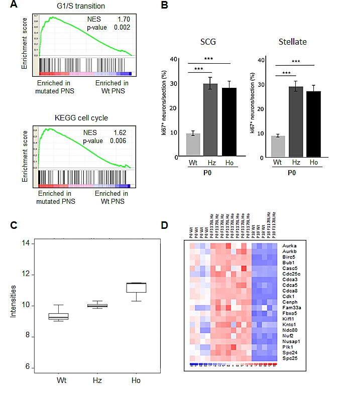
Figure 3. Increased proliferation of sympathetic neurons at P0 in AlkF1178L KI mice.

(A) GSEA of cell cycle and G1/S transition pathway genes in transcriptomic data of Wt and mutated ganglia at P0. The normalized enrichment score (NES) and the nominal p-value are indicated. (B) Quantification of double positive cells for islet1 and ki67 revealed an increased proliferation at P0 in ganglia of heterozygotes (Hz) and homozygotes (Ho) KI AlkF1178L mice. Bonferroni Multiple Comparisons Tests were used to evaluate differences between the groups (n=4 samples in each group). Error bars represent standard deviation. (C) Expression of the Ret gene in sympathetic ganglia at birth. (D) Heatmap of genes belonging to the “Mitosis” category (GO:0007067) in P0 and P18 mutated and Wt ganglia.
