
Figure 6. CD93 silencing affects cell proliferation, adhesion, migration, and in vitro sprouting of human endothelial cells.
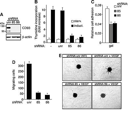
HUVEC were infected with a lentiviral vector expressing unrelated (unr) or CD93 shRNAs (clone 85 or 86). A: Cell extracts from shRNA expressing HUVEC were analyzed by Western blotting using anti-CD93 (H190) and anti-β-actin antibodies to confirm equal loading. B: Cell proliferation expressed as thymidine uptake in infected HUVEC. Cells were grown in 96-well-plates, serum starved (starv) and induced with complete medium (induct). Not infected endothelial cells were also analyzed (-). C: Cell adhesion assay of infected HUVEC. Cells were biochemically detached and allowed to adhere on gelatin-coated surfaces for 15 min. Fixed cells were stained with crystal violet solution and the optical density values were expressed as relative cell adhesion. D: Migration assay on infected HUVEC. Cells were grown in growth factor-depleted culture medium and plated in Boyden chambers. Chemotaxis was stimulated with 10 ng/ml VEGF. Migratory cells were stained and counted under a light microscope. E: Sprouting assay of infected endothelial cells embedded into collagen gels in the absence (NT) or presence of 10 ng/ml VEGF (VEGF). A representative experiment is shown (original magnification, x40). Data represent the ± SD of four-five independent experiments each in triplicate.