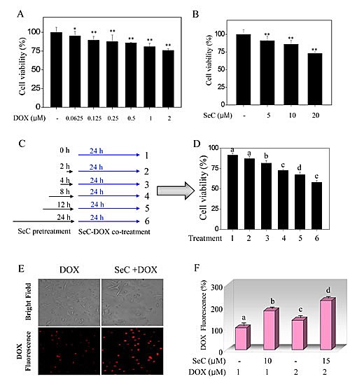
Figure 1. SeC enhances DOX-induced growth inhibition against HepG2 cells through enhanced intracellular uptake of DOX.

Cytotoxic effect of DOX (A) and SeC (B) on HepG2 cells. HepG2 cells (2×10³ cells/well) were seeded in 96-well plate and pre-incubated for 24 h. After incubation, cells were treated with indicated concentration of DOX for 24 h or SeC for 48 h. Cell viability was detected by MTT assay. (C, D) Combined treatment enhanced growth inhibition against HepG2 cells induced by SeC and DOX. The cells were pre-treated with 10 μM SeC (0, 2, 4, 8, 12 and 24 h) and co-incubated with 100 nM DOX for 24 h. Cell viability was determined by MTT assay. Intracellular uptake of DOX was detected by fluorescence microscope (C) and fluorescence micro-plate reader (D). Cells were pre-treated with SeC 24 h and/or DOX for 24 h and assayed by fluorescence microscope (Magnification, 100×) and fluorescence micro-plate reader as described in section of methods. Bars with different characters are statistically different at P<0.05 level.
