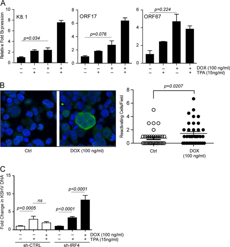
Fig. 4.
Loss of IRF4 results in a robust induction of KSHV lytic gene expression and viral reactivation. (A) qRT-PCR analysis of late genes K8.1, ORF17, and ORF67 in Dox treated BCBL-1 sh-CTRL and sh-IRF4 cells. Cells were stimulated as previously described, total RNA was harvested and subjected to qRT-PCR. Samples were normalized to RPL32 and expressed as fold change with respect to their respective untreated cells (value 1). (B) BCBL-1 sh-IRF4 stimulated with Dox for 96 h. Following stimulation, cells were fixed, stained with KSHV positive human sera (green), nuclei were stained with DAPI (blue), and samples imaged by confocal microscopy. Sample immunofluorescence image is shown (left). Quantification of KSHV reactivation (right). Cells positive for KSHV reactivation were manually counted from a series of ~40 fields. (C) KSHV virion production following IRF4 silencing. BCBL-1 sh-CTRL and sh-IRF4 cells were stimulated with Dox for 72 h followed by stimulation with TPA (15 ng/ml) or Dox and TPA for an additional 72 h. KSHV DNA released into the supernatant was measured by probed-based qPCR. Results are expressed as fold change with respect to their respective untreated cells (value 1).

