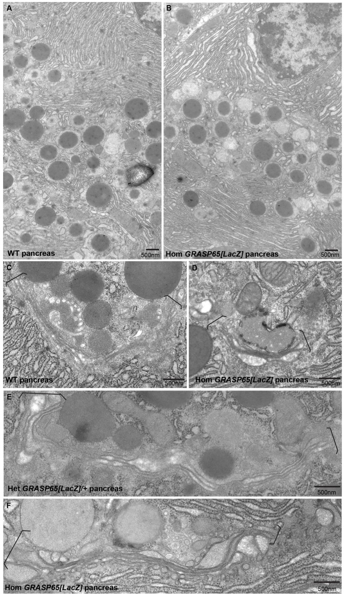
Fig. 5. The Golgi apparatus appears normal in pancreas from GRASP65[LacZ] homozygous mice.
(A,B) Low magnification view of exocrine pancreas in ultrathin epon sections from WT (A) and GRASP65[LacZ] homozygous (B) mice processed for conventional EM. (C–F) High magnification view of Golgi profiles (between brackets) in ultrathin epon sections of exocrine pancreas from WT (A), GRASP65[LacZ] het (C) and hom (D,F) mice. Note that no differences are detectable. Scale bars: 500 nm.

