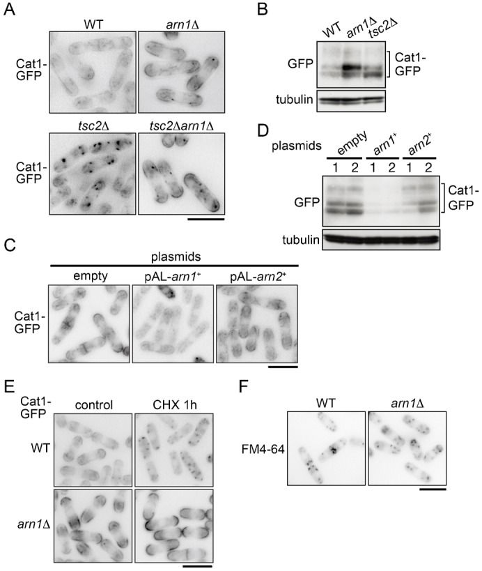
Fig. 3. Arn1 participates in the regulation of the subcellular localization and stability of Cat1.

(A) PJ390 (WT), AN0223 (arn1Δ), PJ380 (tsc2Δ), and AN0224 (tsc2Δarn1Δ) were grown in EMM. (B) PJ390 (WT), AN0223 (arn1Δ), and PJ380 (tsc2Δ) were grown in EMM. Tubulin is shown as a loading control. (C) AN0276 carrying the indicated plasmids was grown in EMM. (D) Cells as in panel C were grown in EMM. (B,D) Proteins were probed with the indicated antibodies. (E) PJ390 (WT) and AN0223 (arn1Δ) were grown in EMM and then treated with 100 µg/ml cycloheximide for 1 hour. (F) PJ390 (WT) and AN0223 (arn1Δ) were grown in EMM and labeled with 50 µM FM4-64 dye on ice for 30 minutes. Five minutes after changing to a fresh medium without the dye at 30°C, the fluorescent signal was observed under a fluorescence microscope. (A,C,E,F) The fluorescence images are shown inverted for clarity. Scale bars: 10 µm.
