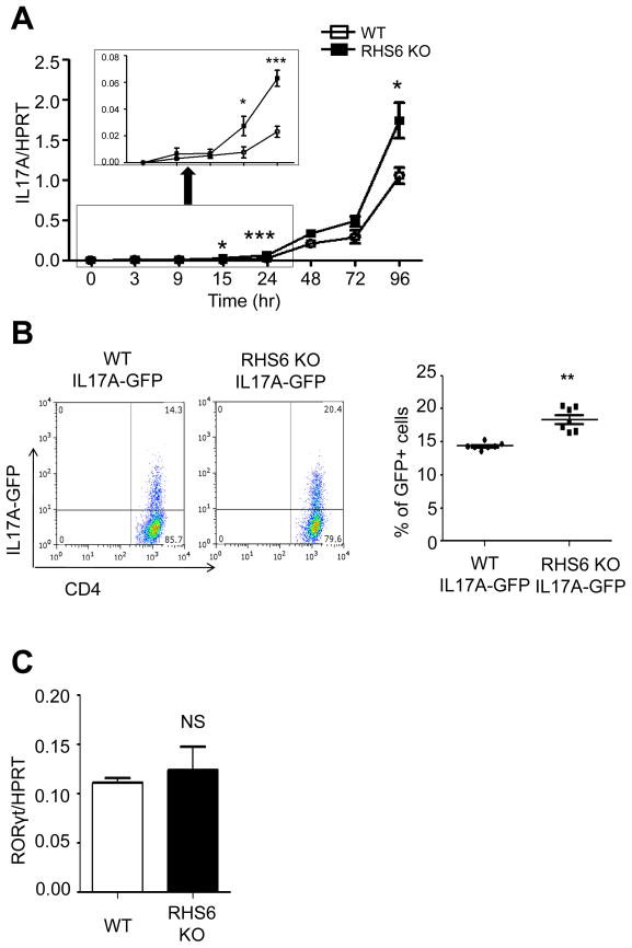
Figure 7. Analysis of IL-17 expression in RHS6 WT and KO T cells.
(A) Quantitative PCR of IL-17 mRNA expression. Naïve T cells from WT and RHS6 KO mice were treated with plate-bound anti-CD3, anti-CD28, TGF-β, IL-6 and IL-23 for the indicated time. The Student t-test was performed (*P < 0.05. ***P < 0.001). (B) MACS purified CD4+ cells from IL-17A-IRES-GFP reporter mice and RHS6 KO IL-17A-IRES-GFP reporter mice were cultured in Th17 polarizing conditions for 2 days. The statistical significance was calculated using the Student t-test (**P < 0.01). (C) Quantitative PCR for RORγt mRNA expression at 24hrs of differentiation. Data are representative of at least three independent experiments and represented as mean +/− SEM. The Student t-test was performed. See also Figure S6.

