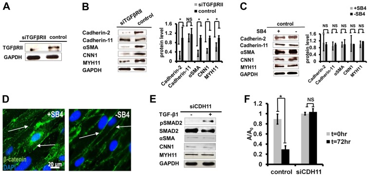
Fig. 5.
High-density-induced SMC differentiation is dependent on TGFβRII signaling. (A,B) siTGFβRII cells were seeded at high-density and, 5 days later, they were lysed. (A) Western blot for TGFβRII. (B) Western blots for cadherin-2, cadherin-11, αSMA, CNN1 and MYH11 (left panel) were quantified to show relative protein levels (right panel). (C) HF-MSCs were seeded at 3×10⁴ cells/cm2 and were treated with SB4 (10 µM) for 5 days. Western blots for cadherin-2, cadherin-11, αSMA, CNN1 and MYH11 (left panel) were quantified to show relative protein levels (right panel). (D) Immunostaining for β-catenin (green). Nuclei were counterstained with DAPI (blue). Arrows indicate adherens junctions. Scale bar: 20 µm. (E) siCDH11 cells were treated with TGF-β1 for 3 days or remained untreated, and the indicated proteins were measured by western blotting. (F) Fibrin hydrogel compaction with siCDH11 or control cells in the presence of exogenous TGF-β1 (10 ng/ml). At each time-point, the area of each gel (‘A’) was measured using ImageJ software and normalized to the initial area (A0). All quantitative data are shown as the mean±s.d.; *P<0.05; NS, not significant.

