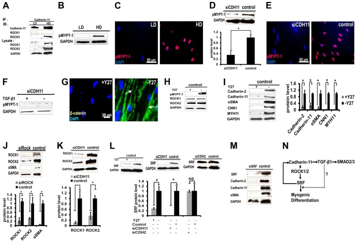
Fig. 6.
Rock and SRF are activated downstream of cadherin-11 and are necessary for high-density-induced myogenic differentiation. HF-MSCs were seeded at 3×10³ (low density, LD) or 30×103 cells/cm2 (high density, HD). (A) Lysates were immunoprecipitated with antibody against cadherin-11. Immunoprecipitates (IP) and corresponding lysates were probed by western blotting (IB). (B) Western blot for pMYPT-1 in cells cultured at low or high density. (C) Immunostaining for pMYPT-1 (pink). Scale bar: 50 µm. (D) Western blot for pMYPT-1 in siCDH11 or control HF-MSCs (upper panel) was quantified to show relative protein levels (lower panel). (E) Immunostaining of siCDH11 or control cells for pMYPT-1 (pink). Scale bar: 50 µm. (F) siCDH11 cells were treated with TGF-β1 (10 ng/ml) and pMYPT-1 was detected by western blotting 3 days later. (G–I) HF-MSCs were seeded at 20×103 cells/cm2, and on day 3 they were treated with Y27 (20 µM) for 1 day. (G) Immunostaining for β-catenin (green). Arrows indicate adherens junctions. Scale bar: 50 µm. In C, E and G, nuclei were counterstained with DAPI (blue). (H) Western blots for pMYPT1, ROCK1 and ROCK2 in Y27-treated and untreated cells. (I) Western blots for cadherin-2, cadherin-11, αSMA, CNN1 and MYH11 (left panel) were quantified to show relative protein levels (right panel). (J) Western blots for the indicated proteins in siROCK cells (upper panel) were quantified to show relative protein levels (lower panel). (K) The level of ROCK1 and ROCK2 in control and siCDH11 cells (lower panel) was determined from western blots (upper panel). (L) HF-MSCs were seeded at 20×103 cells/cm2 and, on day 3, they were treated with Y27 (20 µM) for 1 day. Western blots for SRF in Y27-treated and non-treated cells, siCDH11 or siCDH2 cells (upper panel) were quantified to show relative protein levels (lower panel). All quantitative data in D and I–L show the mean±s.d.; *P<0.05; NS, not significant. (M) SRF was knocked down using shLVDP. Western blots for the indicated proteins in control and siSRF cells are shown. (N) Schematic illustrating the proposed mechanism of cadherin-11-mediated myogenic differentiation.

