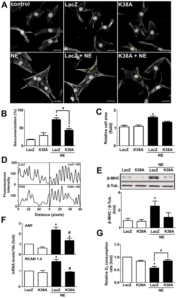
Fig. 4.
Dominant-negative Drp1 (K38A) prevents norepinephrine-induced cardiomyocyte hypertrophy. (A) Representative fluorescence microscopy images of cardiomyocytes transfected with a LacZ- or K38A-encoding adenovirus prior to norepinephrine (NE) stimulation for 48 h, and stained with phalloidin–Rhodamine for sarcomeric detection. Nuclei were stained with Höescht 33342. Scale bar: 20 µm. (B) Percentage of sarcomerized cardiomyocytes and (C) cell area were calculated using at least 55 cells per condition (mean±s.e.m.; n = 4). (D) Fluorescence intensity profile of the yellow lines depicted on A. (E) Western blot analysis of the hypertrophic biomarker β-MHC on cells treated with norepinephrine for 48 h after LacZ or K38A adenovirus transduction (mean±s.e.m.; n = 3). (F) Determination of mRNA levels of atrial natriuretic factor (ANF) and RCAN 1.4 using qPCR (mean±s.e.m.; n = 3). (G) Basal oxygen consumption of Drp1-K38A-transduced cardiomyocytes and stimulated with norepinephrine for 48 h (mean±s.e.m.; n = 4). *P<0.05 versus non stimulated control; #P<0.05 versus norepinephrine-stimulated control.

