
Fig. 4.
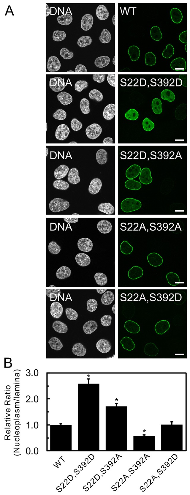
The combined effects of double mutations on the subcellular localization of lamin A. (A) The subcellular distribution of wild-type (WT) GFP–lamin A and the double mutants GFP–lamin A S22D S392D, S22D S392A, S22A S392A and S22A S392D were compared and analyzed as in Fig. 2. GFP–lamin A S22D S392D and S22D S392A accumulated in the nucleoplasm, whereas WT GFP–lamin A, S22A S392A and S22A S392D were, predominantly, localized to the lamina. DNA was stained with Hoechst 33258 and is shown in white. Scale bars: 10 µm. (B) The fluorescence intensities of the lamina and nucleoplasm were quantified, and the relative average ratios of the signals in the nucleoplasm to the signals in the lamina were plotted as in Fig. 2B (see Materials and Methods). Error bars represent the s.e.m. *P<0.005 compared with that of WT GFP–lamin A.