
Figure 4.
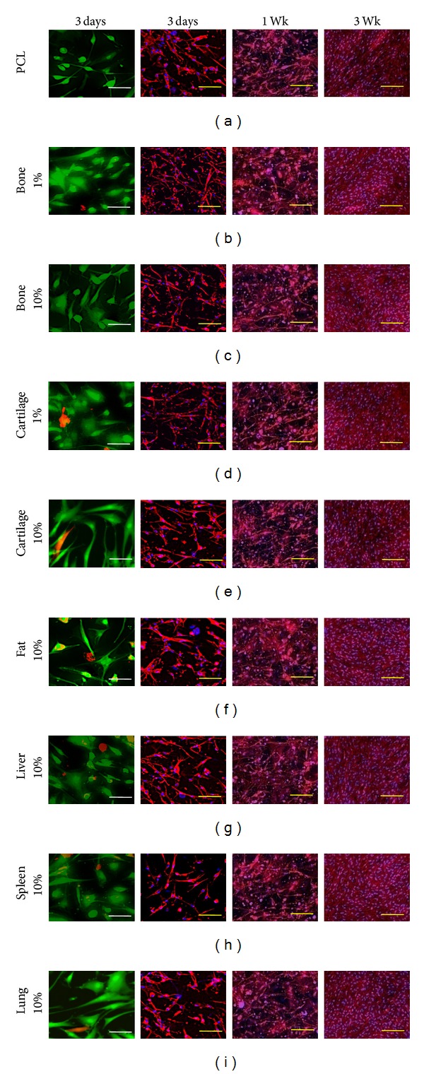
Viability and morphological structure of hASCs grown in monolayer on fibrous scaffolds containing ECM derived from the indicated tissues. In the left panel viable cells are stained green and dead cells are stained red with live/dead staining kit. In the middle and right panels actin is stained red (phalloidin, Texas Red) and cell nucleus is stained blue (DAPI). Scale bars for representative viability and morphology are 20 μm and 100 μm, respectively.