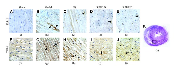
Figure 3.

Immunohistochemical analysis of Toll-like receptors in myocardial tissue. TLR-2 (a–e) and TLR-4 (f–j) expression in early VR rats was observed in each experiment group after AMI using a 400x optical microscopy. Expression of TLRs is much stronger in the tissue of the model group in comparison to the sham group as shown by darker and lager granules (arrowheads in (b) and (g)). The FS and both SNT treatment groups showed reduced expression of TLR-2 and TLR-4 compared to the model group (arrows in (c), (d), (e), (h), (i), and (j)).
